BioImaging
BioImaging (BIM)
BioImaging is an exciting and growing field overlapping the interfaces of engineering, mathematics and computer science, as well as chemistry, physics, life science, and medicine. The main goal of bioimaging is to improve human health by using imaging modalities to advance diagnosis, treatment, and prevention of human diseases.
The BIM track offers high-level interdisciplinary education and training supported by the complementary skills of Université Paris Cité and Arts et Métiers Institute of Technology. A large network of research laboratories provides students access to industrial and experimental imaging systems that utilize innovative technologies.
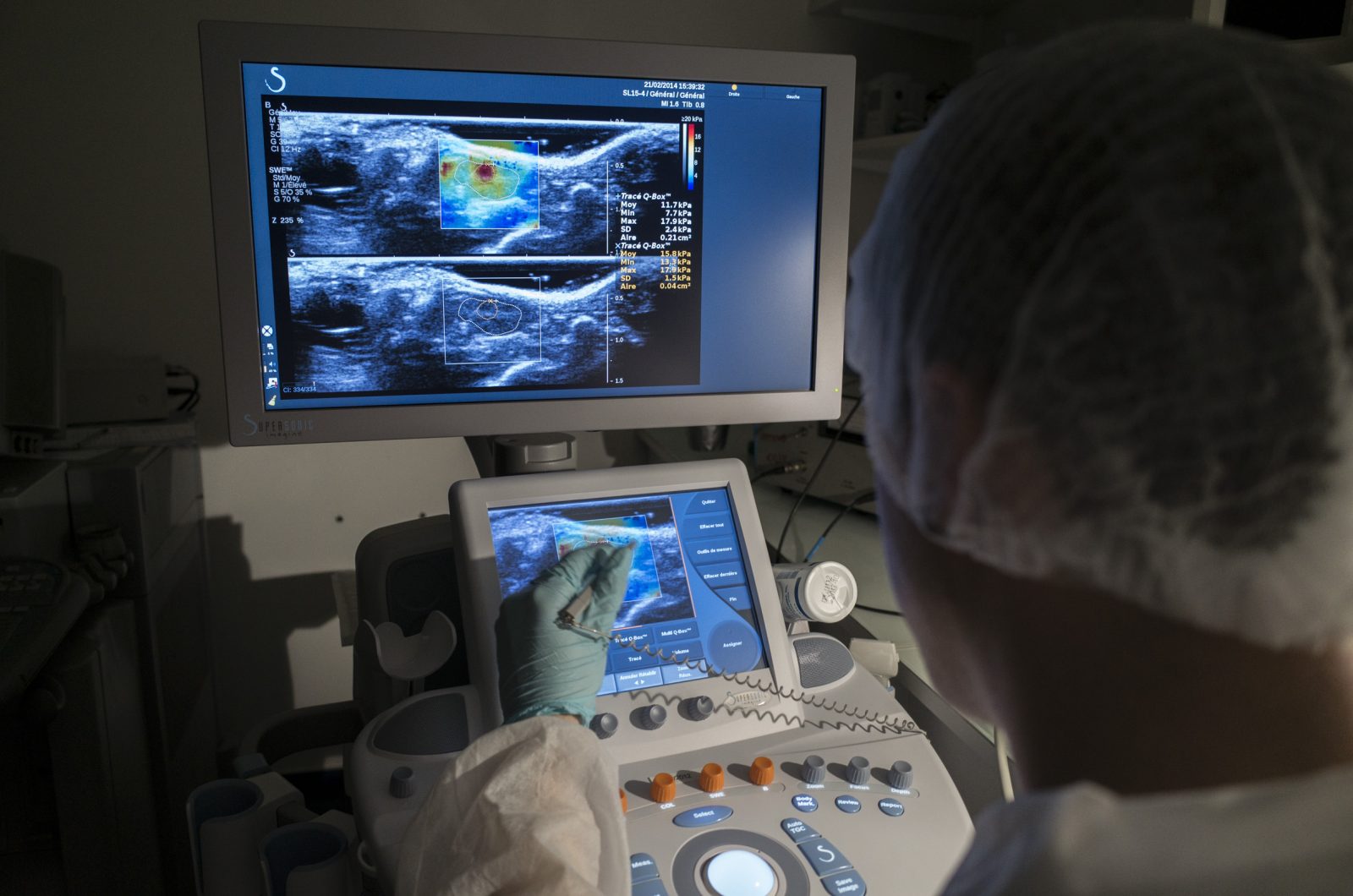
The BIM track is accessible to engineering and life-science students (medicine, pharmacology, biology, chemistry, biochemistry, physics) preparing for career paths in academic research or industrial R&D environments. It relies on close collaboration between Université Paris Cité, Arts et Métiers Institute of Technology in partnership with Telecom Paris.
The third semester (S3) of the BIM curriculum itself consists of six courses (UE) at the M2 level that are organized, taught, and overseen by faculty members expert in the appropriate fields.
The fourth semester (S4) is mainly devoted to complementary skills and the internship (30 ECTS).
Semester 3
Mandatory courses
Course Title: Open Your Mind Seminars
Description:
Key words:
Total number of hours: Number of ECTS: Semester
Mandatory course ☐ Optional course ☐
Prerequisites/skills needed:
Teaching methods and activities:
Location:
Course supervisor:
Course Title: Interdisciplinary week
Description:
Key words:
Total number of hours: Number of ECTS: Semester
Mandatory course ☐ Optional course ☐
Prerequisites/skills needed:
Teaching methods and activities:
Location:
Course supervisor:
Course Title: Medical Image Analysis
Description: The main objective of this course is to provide the students with the means to understand and use the most common tools used in bio-medical image analysis, this will include notions about the sampling and quantification of images, image filtering, image segmentation, Image segmentation, video sequences & motion processing, 3-D Image processing, and short introduction to the extraction/measurement of quantitative features and pattern recognition.
Key words: Image processing, Bio-medical images
Total number of hours: 40 Number of ECTS: 6 Semester 3
Mandatory course ☒ Optional course ☐
Prerequisites/skills needed: This course requires familiarity with basic mathematics: Linear algebra: linear equations, vectors, matrices; Analysis: continuous function, graph of a function, basic notions of differentiation and integration; Geometry: point, segment, line, curve, surface; Probabilities: probability distribution. Engineers are expected to have some basic knowledge in programming with Matlab, Java or C/C++. Basic knowledge about numerical analysis is also expected
Teaching methods and activities: Lectures (CM), practical sessions (TD), other: project
Location: Arts et Métiers ParisTech
Course supervisor: Florence CLOPPET (associate professor), Petr DOKLADAL (engineer) and Nicolas LOMENIE (associate professor)
One course (6 ECTS) to be picked among
Course Title: Physics for BioImaging
Description: Physical principles behind biomedical imaging technologies. Beginning with the fundamental principles for X-ray computed tomography, magnetic resonance imaging, nuclear medicine and ultrasound students will learn the physical concepts related to detectors and image formation. Principles are related to practical imaging configurations so that knowledge can be applied to better select and design imaging toward new applications. Most recent advances in biomedical imaging technologies such as novel detectors and multi-wave techniques are presented by expert researchers to prepare for the biomedical imaging environment of the future. Ultrasound physics for wave propagation and scattering are explained and further developed to include principles behind multiwave imaging and ultrasound therapy. Physics principles behind the detection of ultrasound contrast agents are covered. Physics of ionizing radiation is explained PET Gamma Cameras and PET scans are explained along with the basic physics of computed tomography. New flat panel detectors for conventional radiology and strategies for dose reduction and other new perspectives are presented. . MRI principles and diffusion and spectrometry MRI are explained (Means to use MRI to evaluate microcirculation and inhomogeneous Magnetization transfer). Imaging-guided therapies are increasingly used, replacing surgery in certain situations. Simulator work will allow the student to become familiar with these techniques.
Key words: MRI, CT, Nuclear Medicine, Ultrasound, Physics, image formation, instrumentation
Total number of hours: 40 Number of ECTS: 6 Semester 3
Mandatory course ☐ Optional course ☒
Prerequisites/skills needed: Principles of Fourier Transform, Physics notions: electromagnetism, magnetostatics, spin magnetic moments, radiation, wave propagation
Teaching methods and activities: Lectures (CM), practical sessions (TD), other: visits on platforms
Location: Université Paris Descartes, Centre Universitaire des Saints Pères
Course supervisor: Lori BRIDAL (researcher) and Mathilde WAGNER (associate professor)
Course Title: Chemistry for Bioimaging : Basics, probes and nanomedicine
Description: The chemistry for bioimaging course (innovative probes and nanomedicine) is dedicated to engineers, physicists, physicians, biologists, chemists. It brings expertise in chemistry and chemical properties of imaging probes used and developed in medical and biomedical imaging, in particular molecular and functional imaging using chemical imaging agents. This knowledge and expertise in the chemical properties of imaging agents will allow users to know how to use them in practice wisely for diagnostic purposes for the detection of pathologies as well as the monitoring of therapies. This teaching will allow to know how to manage their preparation, their possible formulation and their route of administration, associated with the methods of acquisition of imaging in all the modalities (MRI, Ultrasound, Optics, Nuclear Medicine, EPR, CT). It will inversely supply the knowledge of the commercial and innovative imaging agents to be able to perform a better preclinical or clinical diagnosis, with existing and developing medical imaging modalities. The latest development of imaging probes will be taught, and illustrated with many recent applications in preclinical and clinical. In addition, practical course demonstrations on all In Vivo imaging facilities of Paris Descartes, scanning all imaging modalities, will be organized in order to understand concretely the applications.
Key words: Innovative Probes, Nanomedicine, Pre-clinical and Clinical Diagnosis
Total number of hours: 45 Number of ECTS: 6 Semester 3
Mandatory course ☐ Optional course ☒
No prerequisites/skills needed
Teaching methods and activities: Lab sessions (TP), lectures (CM), other: visits on platforms
Location: Université Paris Descartes, Centre Universitaire des Saints Pères
Course supervisor: Bich-Tuy DOAN (researcher) and Yves FRAPART (researcher ingineer)
Course Title: Optical Imaging
Description: The objective of the course is to give insights into modern research trends in biophotonics. The course provides in particular an overview of the various optical techniques available for biomedical imaging and detection, giving their characteristics and highlighting their advantages and drawbacks.From widefield microscopy to super-resolution and two-photon imaging, this course of 36h alternating theory lectures and practical labworks will provide the essential notions in modern light microscopy. New techniques are discussed: widefield, confocal, non-linear microscopy, super-resolution approaches (PALM, STORM, STED, SIM). From single protein and simple cell layer to the whole organ, model organisms and small animals, we will discuss about the most adapted imaging technique. Notions of image analysis will be presented, either on 3D visualization or on deconvolving and filtering the images acquired during the labworks.
Key words: Light microscopy, image processing, linear optics, non-linear optics, super-resolution microscopy
Total number of hours: 36 Number of ECTS: 6 Semester 3
Mandatory course ☐ Optional course ☒
Prerequisites/skills needed: Linear optics, good notions in biology
Teaching methods and activities: Lab sessions (TP), lectures (CM)
Location: Université Paris Descartes, Centre Universitaire des Saints Pères and Institut Cochin
Course supervisor: Benoit FORGET (professor) and Thomas GUILBERT (researcher ingineer)
15 ECTS (minimum) to be picked among
Course Title: Molecular Imaging
Description: Molecular Imaging is non invasive in vivo biochemistry: in vivo means real-life environment, non invasive means repeatable, molecular biology (~biochemistry) means relevant to molecular diseases such as cancer, inflammation, cardiovascular diseases, etc. The course will present practical up-to-date examples of Whole body, 3D, Quantitative, Dynamic and Multiplexed molecular imaging research and achievements.
Key words: Molecular imaging, molecular medicine, experimental imaging
Total number of hours: 20 Number of ECTS: 3 Semester 3
Mandatory course ☐ Optional course ☒
Prerequisites/skills needed: Interest for Molecular aspects of life
Teaching methods and activities: Lectures (CM), other: personal assignment
Location: Université Paris Descartes, Centre Universitaire des Saints Pères
Course supervisor: Bertrand TAVITIAN (professor) and François ROUZET (professor)
Course Title: Functional and Metabolic Imaging
Description: This course provides a general introduction to functional and metabolism imaging techniques (fMRI, Diffusion MRI and PET) and concrete examples of clinical applications with hands-on experience of processing data on clinical routine workstations. It is composed of 2 parts organized as follows: 1. Brain Imaging: General introduction of Imaging techniques and associated clinical applications (PET, – Functional neuroimaging, Diffusion-weighted magnetic resonance imaging) and Hands-on on Image analysis of clinical cases on routine workstations (Perfusion MRI and PET: multimodality fusion on several clinical cases, Diffusion imaging and fMRI: Application to brain tumors) 2. Body Imaging: Introduction to the concept of functional and metabolism imaging (MRI, CT and ultrasound), Imaging of MRI diffusion, Quantitative imaging of microcirculation (A hands-on session is proposed using a digital microcirculation simulator), Imaging of tissue elasticity (Elastography) by MRI and ultrasound. Radiomics (principles and methodology): the links with other -omics are explained and the relationship with the phenotype is explored. Notions illustrated by applications related to different organs: liver, ENT organs, kidneys … and cancerous tumors. The applications related to the prognosis of the lesions and the evaluation under treatment are also described.
Key words: Brain Imaging, Body Imaging, MRI Diffusion, Elastography, Radiomics, Multimodality
Total number of hours: 24 Number of ECTS: 3 Semester 3
Mandatory course ☐ Optional course ☒
No prerequisites/skills needed
Teaching methods and activities: Lectures (CM), practical sessions (TD)
Location: Université Paris Descartes, Centre Universitaire des Saints Pères
Course supervisor: Charles-André CUENOD (PU-PH) and Myriam EDJALI-GOUJON (doctor)
Course Title: Quantification for Diagnosis: Industrial and Medical Applications in Medical Image Analysis
Description: The objective of the course is to show the students a panorama of several clinical and industrial applications in medical image analysis. The course will be taught by pairs of academic or industrial researchers and clinicians working together on the same clinical problem. They will show how mathematical models and algorithms can answer to clinical demands producing tools, patents and machines used on a daily basis in hospitals.
Key words: Industrial medical applications, clinical applications
Total number of hours: 24 Number of ECTS: 3 Semester 3
Mandatory course ☐ Optional course ☒
Prerequisites/skills needed: Industrial medical applications, clinical applications
Teaching methods and activities: Lectures (CM), practical sessions (TD), other: personal assignment (bibliography report)
Location: Arts et Métiers ParisTech
Course supervisor: Pietro GORI (associate professor) and Elsa ANGELINI (associate professor)
Course Title: Quantification for Neuroimaging
Description: The module aims at providing students with an insight on advanced methods in data modelling and analysis as used in neuroimaging research labs. The methods are selected to present four differents aspects of brain acitivity and connectivity: – diffusion MRI and tractography – activation fMRI and activation networks – resting state fMRI and resting state connectivity – Magneto Encephalography and source reconstruction Each lecture starts with a presentation of the critical theoretical knowledge regarding the neuroimaging modality, the specificities of the generated data, and the methodology for image analysis. Then, praticals on state-of-the-art softwares and real datasets provide hands-on experience of data processing. This interplay between theory and practice teaches students how to apply their knowledge and skills to handle cutting-edge methods applied to neuroimaging data and how to consider the limitations of such methods and their impact on the interpretation of results. Critically, the lectures present various methodological developments to overcome these issues (artifact correction, bias modeling,…).
Key words: Functional MRI, Electroencephalography, Magnetoencephalography
Total number of hours: 20 Number of ECTS: 3 Semester 3
Mandatory course ☐ Optional course ☒
Prerequisites/skills needed: Linear algebra, Basic programming skills (Matlab or Python)
Teaching methods and activities: Lectures (CM), practical sessions (TD), other: personal assignment
Location: Arts et Métiers ParisTech
Course supervisor: Alexandre GRAMFORT (researcher) and Sylvain CHARRON (researcher engineer)
Course Title: Quantification for Bioimaging
Description: This lecture provides basic knowledge about the key-concepts of the bioimaging analysis from optical microscopy. It is composed of 5 classes divided in a theorical part and pratical part by manipulating images under the Icy platform : Part 1: General overview of the bioimaging analysis and basic concepts: Part 2: Spot detection and tracking (intra-cellular particles) Labeling intra-cellular particles (virus, endosomes, etc.) can be seen as spots in noisy background. We will present the approaches of Part 3: Cell detection and tracking from video microscopy sequences (deformable models) Presentation of the interest of the detection and analysis from video microscopy sequences Part 4: Digital pathology (color representation, color image processing, big data).
Key words: Spot Detection, Cell Detection, Tracking, Video-microscopy, Digital Pathology
Total number of hours: 20 Number of ECTS: 3 Semester 3
Mandatory course ☐ Optional course ☒
No prerequisites/skills needed.
Teaching methods and activities: Lectures (CM), practical sessions (TD), other: personal assignment
Location: Arts et Métiers ParisTech
Course supervisor: Vannary MEAS-YEDID (researcher engineer)
Course Title: Machine Learning
Description: – Learn the principles and hypothesis behind the different machine learning techniques – Understand the pros and cons of every method – Learn how to use them on real high-dimensional biomedical imaging data (Big Data) – Use different techniques in practice.
Key words: Statistical machine learning, deep learning
Total number of hours: 23 Number of ECTS: 3 Semester 3
Mandatory course ☐ Optional course ☒
Prerequisites/skills needed: Basic courses in Linear Algebra, Statistics, Optimization and Programming (for clinicians this is not mandatory but they must be willing to code and see equations during the lectures …)
Teaching methods and activities: Lectures (CM), practical sessions (TD)
Location: Arts et Métiers ParisTech
Course supervisor: Pietro GORI (associate professor) and Petr DOKLADAL (associate professor)
Course Title: Physics for BioImaging
Description: Physical principles behind biomedical imaging technologies. Beginning with the fundamental principles for X-ray computed tomography, magnetic resonance imaging, nuclear medicine and ultrasound students will learn the physical concepts related to detectors and image formation. Principles are related to practical imaging configurations so that knowledge can be applied to better select and design imaging toward new applications. Most recent advances in biomedical imaging technologies such as novel detectors and multi-wave techniques are presented by expert researchers to prepare for the biomedical imaging environment of the future. Ultrasound physics for wave propagation and scattering are explained and further developed to include principles behind multiwave imaging and ultrasound therapy. Physics principles behind the detection of ultrasound contrast agents are covered. Physics of ionizing radiation is explained PET Gamma Cameras and PET scans are explained along with the basic physics of computed tomography. New flat panel detectors for conventional radiology and strategies for dose reduction and other new perspectives are presented. . MRI principles and diffusion and spectrometry MRI are explained (Means to use MRI to evaluate microcirculation and inhomogeneous Magnetization transfer). Imaging-guided therapies are increasingly used, replacing surgery in certain situations. Simulator work will allow the student to become familiar with these techniques.
Key words: MRI, CT, Nuclear Medicine, Ultrasound, Physics, image formation, instrumentation
Total number of hours: 40 Number of ECTS: 6 Semester 3
Mandatory course ☐ Optional course ☒
Prerequisites/skills needed: Principles of Fourier Transform, Physics notions: electromagnetism, magnetostatics, spin magnetic moments, radiation, wave propagation
Teaching methods and activities: Lectures (CM), practical sessions (TD), other: visits on platforms
Location: Université Paris Descartes, Centre Universitaire des Saints Pères
Course supervisor: Lori BRIDAL (researcher) and Mathilde WAGNER (associate professor)
Course Title: Chemistry for Bioimaging : Basics, probes and nanomedicine
Description: The chemistry for bioimaging course (innovative probes and nanomedicine) is dedicated to engineers, physicists, physicians, biologists, chemists. It brings expertise in chemistry and chemical properties of imaging probes used and developed in medical and biomedical imaging, in particular molecular and functional imaging using chemical imaging agents. This knowledge and expertise in the chemical properties of imaging agents will allow users to know how to use them in practice wisely for diagnostic purposes for the detection of pathologies as well as the monitoring of therapies. This teaching will allow to know how to manage their preparation, their possible formulation and their route of administration, associated with the methods of acquisition of imaging in all the modalities (MRI, Ultrasound, Optics, Nuclear Medicine, EPR, CT). It will inversely supply the knowledge of the commercial and innovative imaging agents to be able to perform a better preclinical or clinical diagnosis, with existing and developing medical imaging modalities. The latest development of imaging probes will be taught, and illustrated with many recent applications in preclinical and clinical. In addition, practical course demonstrations on all In Vivo imaging facilities of Paris Descartes, scanning all imaging modalities, will be organized in order to understand concretely the applications.
Key words: Innovative Probes, Nanomedicine, Pre-clinical and Clinical Diagnosis
Total number of hours: 45 Number of ECTS: 6 Semester 3
Mandatory course ☐ Optional course ☒
No prerequisites/skills needed
Teaching methods and activities: Lab sessions (TP), lectures (CM), other: visits on platforms
Location: Université Paris Descartes, Centre Universitaire des Saints Pères
Course supervisor: Bich-Tuy DOAN (researcher) and Yves FRAPART (researcher ingineer)
Course Title: Optical Imaging
Description: The objective of the course is to give insights into modern research trends in biophotonics. The course provides in particular an overview of the various optical techniques available for biomedical imaging and detection, giving their characteristics and highlighting their advantages and drawbacks.From widefield microscopy to super-resolution and two-photon imaging, this course of 36h alternating theory lectures and practical labworks will provide the essential notions in modern light microscopy. New techniques are discussed: widefield, confocal, non-linear microscopy, super-resolution approaches (PALM, STORM, STED, SIM). From single protein and simple cell layer to the whole organ, model organisms and small animals, we will discuss about the most adapted imaging technique. Notions of image analysis will be presented, either on 3D visualization or on deconvolving and filtering the images acquired during the labworks.
Key words: Light microscopy, image processing, linear optics, non-linear optics, super-resolution microscopy
Total number of hours: 36 Number of ECTS: 6 Semester 3
Mandatory course ☐ Optional course ☒
Prerequisites/skills needed: Linear optics, good notions in biology
Teaching methods and activities: Lab sessions (TP), lectures (CM)
Location: Université Paris Descartes, Centre Universitaire des Saints Pères and Institut Cochin
Course supervisor: Benoit FORGET (professor) and Thomas GUILBERT (researcher ingineer)
Course Title: Advanced Optical Imaging
Description:
Key words:
Total number of hours: Number of ECTS: Semester
Mandatory course ☐ Optional course ☐
Prerequisites/skills needed:
Teaching methods and activities: l
Location:
Course supervisor:
During the S3, students select five 6-ECTS courses (UE). The set of each student’s elective courses is determined according to his/her professional objectives and approved by their track advisor.
Semester 4
Course Title: Ethical and Industrial Aspects
Description:
Key words:
Total number of hours: Number of ECTS: Semester
Mandatory course ☐ Optional course ☐
Prerequisites/skills needed:
Teaching methods and activities:
Location:
Course supervisor:
Course Title: Research Internship
Description:
Key words:
Total number of hours: Number of ECTS: Semester
Mandatory course ☐ Optional course ☐
Prerequisites/skills needed:
Teaching methods and activities:
Location:
Course supervisor:
As far as the non mandatory courses are concerned, the students’ choices will have to be discussed with the track chairs and will be finalized during the interdisciplinary week, taking the student’s personal background and interests into consideration. It will then be validated in a co-signed contract of study.
The courses students will eventually attend will also depend on actual availability: some courses might not open because of an insufficient number of attendees, while others may have a limited capacity and not be able to offer a seat to all.
As indicated, a course may be offered to BME Paris students outside the track which supports it. In such cases, priority will be given to students enrolled in the supporting track.
Students enrolled in other ENSAM, Université Paris Descartes or Université PSL programs may also attend a BME Paris course, provided that:
- their respective program chairs support their application
- the course is not already full
- the relevant BME Paris chairs accept their application which must include which a CV and a motivation letter.
Course Title: A window into the mind : new technologies to explore and stimulate the brain
Description: This UE presents the following topics as 2- to 3h lectures : Overview of the neurophysiological mechanisms related to the different methods of exploration of brain activity – Principles of experimental research in Neuroscience – Exploration of Brain activity: Basic concepts and description of intracranial recordings in behaving animals and optogenetics – Non-invasive exploration and stimulation in human brain: fIRM and TMS – Acoustical brain imaging: from observation to treatment – Optogenetics and machine learning in Neuroscience – Brain imaging with voltage-sensitive dyes – Two-photon microscopy introduction and new evolution: Fast multisite optical recordings with acousto-optic deflectors. The UE also includes one day devoted to presentations of scientific papers by students in front of the class.
Key words: Brain imaging, optogenetics, ultrasound
Total number of hours: 29 Number of ECTS: 3 Semester 3
Mandatory course ☒ Optional course ☒
Prerequisites/skills needed: Some basic neuroscience (cf UE Refresher courses), notions in frequency analysis
Teaching methods and activities: Lectures (CM), other: article presentations by students
Location: ESPCI Paris
Course supervisor: Karim BENCHENANE (researcher)
Course Title: Brain-Computer Interfaces: from modeling to engineering
Description: By the end of this teaching unit, students will be able to design a brain-computer interfaces (BCI) system by themselves. The purpose of the training unit is twofold: (i) to provide students with basic knowledge in signal processing and machine learning, which are ubiquitous tools for the modeling of complex systems such as the brain and neural systems, (ii) to describe actual applications of the concepts presented in the first three courses, especially BCI. A first two courses will present the methodological tools for designing valid data-driven models. Courses 3 to 5 will describe applications to electroencephalographic modeling and BCI. Course 6 will present applications of modeling to spike sorting. Course 7 will present an introduction and applications of deep learning in biomedical engineering. Course 8 will involve team projects (8 x 4h-sessions).
Key words: EEG, machine learning, executive functions, signal processing
Total number of hours: 50 Number of ECTS: 6 Semester 3
Mandatory course ☒ Optional course ☐
Prerequisites/skills needed: Some statistics (cf UE 3.2)
Teaching methods and activities: Lab sessions (TP), lectures (CM), other: reverse learning
Location: ESPCI Paris, La Pitié-Salpétrière hospital
Course supervisor: FrançoisVIALATTE (Associate Professor)
Course Title: Basics in Tissue and Cell Biology
Description: This lecture is for scientific students who didn’t receive a biological/medical knowledge. The classes provide a fundamental knowledge about the basics in biology. The lecture is composed of 4 parts:
Part 1 – General principles of cell and molecular biology:
– Cell structures and molecular components of the cell (nucleic acids, proteins, lipids, sugars)
– Cell proliferation, differentiation and death
– Regulation of gene expression
– Introduction to cell metabolism
– Interactions between the cell and its environment
Part 2 – Basic techniques of cell and molecular biology:
– Cell biology: – cell culture / – microscopy
– Molecular biology: – computer-assisted sequence-analyses (nucleic acids, proteins) /- extraction and analyses of nucleic acids, proteins and of their interactions /- DNA cloning, mutagenesis, transfection
Part 3 – Mechanical stress and cell signaling in cartilage and intervertebral disc
Part 4 – Cartilage cell biology and bioengineering
Key words: Cell biology
Total number of hours: 24 Number of ECTS: 3 Semester 3
Mandatory course ☒ Optional course ☐
No prerequisites/skills needed.
Teaching methods and activities: Lectures (CM)
Location: Arts et Métiers ParisTech
Course supervisor: François RANNOU (PU-PH) and Caroline CHAUVET (associate professor)
Course Title: Practical training in bioengineering
Description:
Key words:
Total number of hours: Number of ECTS: Semester
Mandatory course ☐ Optional course ☐
Prerequisites/skills needed:
Teaching methods and activities:
Location:
Course supervisor:
Course Title: Biosensors for medical diagnosis
Description:
Key words:
Total number of hours: Number of ECTS: Semester
Mandatory course ☐ Optional course ☐
Prerequisites/skills needed:
Teaching methods and activities: :
Location:
Course supervisor:
Course Title: Anatomy of the Musculo-skeletal System
Description: This course provides basics in functional anatomy focusing on the osteoarticular and muscular systems and their links with the movement. 6 anatomical regions are presented: foot and ankle, hip, knee, spine, elbow and wrist, shoulder. Clinical issues in the orthopaedic field such as prosthetic fitting are also presented.
Key words: Functional anatomy, osteoarticular system, muscles
Total number of hours: 24 Number of ECTS: 3 Semester 3
Mandatory course ☒ Optional course ☐
No prerequisites/skills needed.
Teaching methods and activities: Lectures (CM)
Location: Arts et Métiers ParisTech
Course supervisor: Philippe WICART (professor)
Course Title: Research Methodology
Description:
Key words:
Total number of hours: Number of ECTS: Semester
Mandatory course ☐ Optional course ☐
Prerequisites/skills needed:
Teaching methods and activities:
Location:
Course supervisor:

